Microscale imaging sheds light on species‐specific strategies for photo‐regulation and photo‐acclimation of microphytobenthic diatoms
Intertidal microphytobenthic (MPB) biofilms are key sites for coastal primary production, predominantly by pennate diatoms exhibiting photo‐regulation via non‐photochemical quenching (NPQ) and vertical migration. Movement is the main photo‐regulation mechanism of motile (epipelic) diatoms and because they can move from light, they show low‐light acclimation features such as low NPQ levels, as compared to non‐motile (epipsammic) forms. However, most comparisons of MPB species‐specific photo‐regulation have used low light acclimated monocultures, not mimicking environmental conditions. Here we used variable chlorophyll fluorescence imaging, fluorescent labelling in sediment cores and scanning electron microscopy to compare the movement and NPQ responses to light of four epipelic diatom species from a natural MPB biofilm. The diatoms exhibited different species‐specific photo‐regulation features and a large NPQ range, exceeding that reported for epipsammic diatoms. This could allow epipelic species to coexist in compacted light niches of MPB communities. We show that diatom cell orientation within MPB can be modulated by light, where diatoms oriented themselves more perpendicular to the sediment surface under high light vs. more parallel under low light, demonstrating behavioural, photo‐regulatory response by varying their light absorption cross‐section. This highlights the importance of considering species‐specific responses and understanding cell orientation and photo‐behaviour in MPB research.
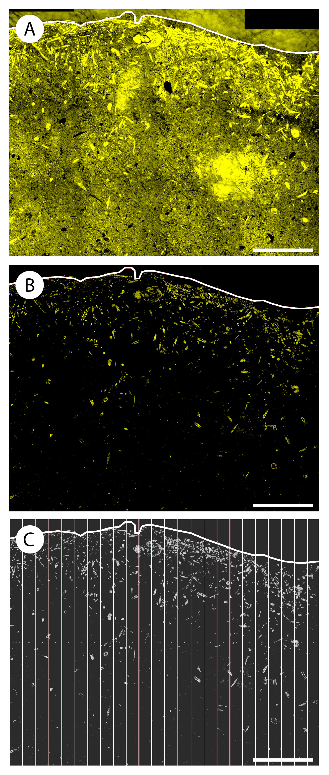
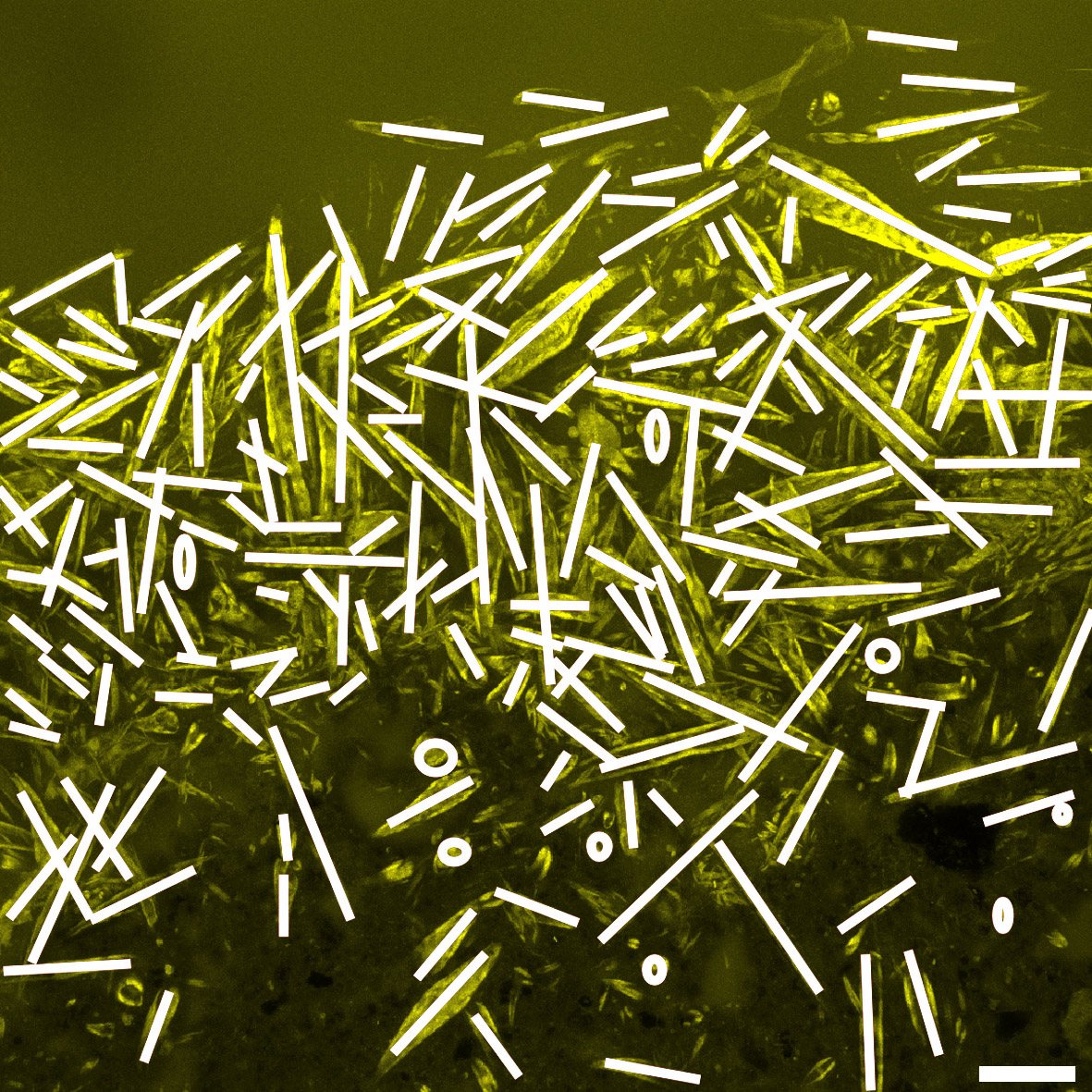
Figure S1. Workflow to quantify diatom pixels in relation to the sediment surface. Example of a portion of a HL sample. (A) FLEC image of a sediment profile used to manually draw the sediment ...
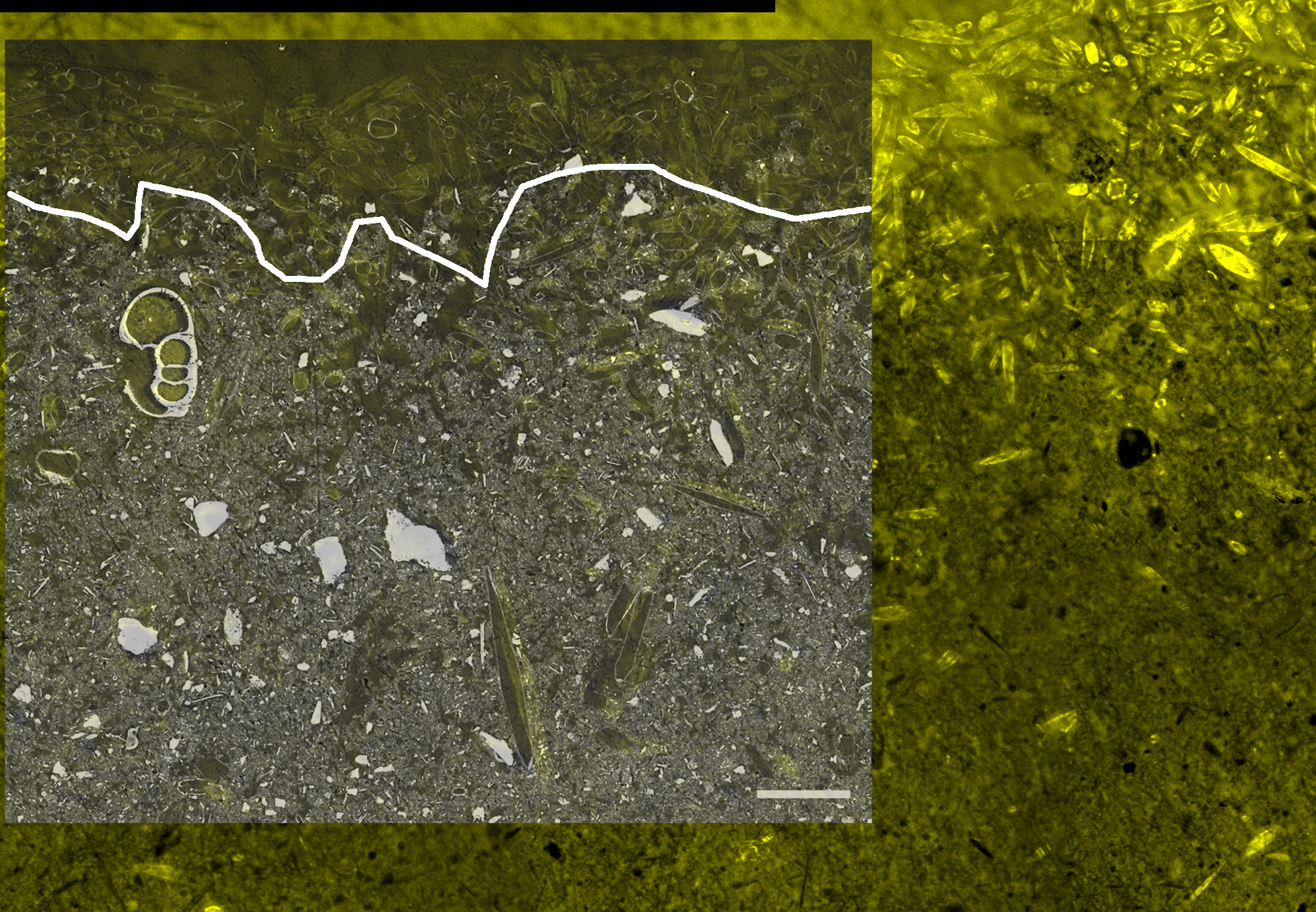
Figure S3. Sediment profile of a LL SEM image overlaid on the corresponding FLEC image. Overlaying these two types of images helped with the detection of the sediment surface. Sediment appears in ...
Jesus Bruno, Jauffrais Thierry, Trampe Erik, Méléder Vona, Ribeiro Lourenço, Bernhard Joan M., Geslin Emmanuelle, Kühl Michael (2023). Microscale imaging sheds light on species‐specific strategies for photo‐regulation and photo‐acclimation of microphytobenthic diatoms. Environmental Microbiology. 25 (12). 3087-3103. https://doi.org/10.1111/1462-2920.16499, https://archimer.ifremer.fr/doc/00853/96459/